Buy or rent our products online!
A concussion is classified as a mild traumatic brain injury (mTBI) — but for the up to 30% of concussion patients who develop post-concussion syndrome (PCS), there is nothing mild about it. Headaches that won’t relent. Brain fog that makes reading feel impossible. Memory gaps, crushing fatigue, light sensitivity, mood swings, and sleep that never feels restorative — sometimes persisting months or years after the original injury.
The most common and frustrating experience for people with PCS is being told by standard neuroimaging — MRI, CT scan — that their brain looks normal. So why do they still feel so impaired? The answer lies in the brain’s electrical activity, not its anatomy. And it is precisely why quantitative EEG (qEEG) brain mapping and qEEG-guided neurofeedback have become the most targeted, evidence-supported tools available for post-concussion recovery.
What Is Post-Concussion Syndrome — and Why Do Symptoms Persist?
A concussion occurs when a bump, jolt, or blow to the head — or a rapid back-and-forth movement of the body — causes the brain to move and twist within the skull. This mechanical force triggers a neurochemical cascade: glutamate floods the synaptic space, potassium ions flow out of neurons, calcium floods in, and the brain’s energy metabolism is severely disrupted. For most people, this resolves within 7–14 days. For those with PCS, it does not.
The emerging neurological explanation for persistent symptoms is disrupted neurovascular coupling — the brain’s mechanism for directing blood flow and oxygen to regions doing active work. After a concussion, the brain reroutes resources around injured sites to still-functioning areas. In PCS, this distribution never normalises: some regions become chronically overloaded, others become chronically underactive. The result is persistent brainwave dysregulation — measurable, reproducible patterns of abnormal electrical activity — that standard MRI and CT cannot detect because they image structure, not function.
This is the critical clinical gap: a patient can have a structurally normal brain on MRI and profoundly abnormal brainwave activity on qEEG. The symptoms are not imagined. They are neurologically real — and they are measurable.
The Full Spectrum of Post-Concussion Symptoms
Post-concussion syndrome produces symptoms across three overlapping domains. The breadth of the symptom profile — and the degree to which different symptom clusters dominate — varies by patient, injury mechanism, and the specific brain regions affected.
| Physical | Cognitive | Emotional / Behavioural |
| Persistent headaches / migraines | Brain fog / mental cloudiness | Anxiety / depression |
| Dizziness / balance problems | Memory problems | Irritability / emotional volatility |
| Visual disturbances | Difficulty concentrating | Personality changes |
| Light & sound sensitivity | Slowed processing speed | Low frustration tolerance |
| Fatigue / sleep disruption | Executive function deficits | Social withdrawal |
| Tinnitus / ringing ears | Word-finding difficulty | PTSD symptoms (in mTBI) |
An important clinical note: symptoms typically emerge or intensify in cognitively demanding situations — reading, screen use, noise, stress, and complex social environments — because these activities push the already-strained dysregulated brain circuits past their limited capacity. This is not a psychological weakness. It is a neurological resource limitation, directly measurable on qEEG.
Why Standard Treatments Often Fall Short
The conventional medical approach to post-concussion syndrome centres on rest, time, and symptom management — analgesics for headache, SSRIs for mood, sleep aids, and cognitive rest protocols. For many patients, these provide partial relief at best. The fundamental limitation is that they address symptoms rather than the underlying brainwave dysregulation driving them.
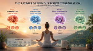
Rest and time allow the brain’s acute neurochemical injury to resolve — but they do not retrain the disrupted brainwave patterns that become established after the initial injury window. Excess slow-wave delta in waking state does not spontaneously normalise through rest alone in PCS patients. Disrupted coherence between brain regions does not repair itself without targeted retraining. This is why patients who receive only rest-based protocols often plateau — they are well past the acute injury phase but their brain’s electrical function has not been retrained toward pre-injury patterns.
What qEEG Reveals That MRI and CT Cannot
Quantitative electroencephalography (qEEG) measures and maps the brain’s electrical activity across 19 electrode sites, processing the data through Fast Fourier Transform (FFT) spectral analysis and comparing the results to an FDA-recognised normative database of neurologically healthy individuals matched by age and gender. In post-concussion patients, this produces a picture of the brain’s actual functional state — the dysregulation that is driving symptoms — that structural imaging simply cannot see.
Typical qEEG Findings in Post-Concussion Syndrome• Excess slow-wave delta (0.5–4 Hz) activity in waking state — the hallmark of cortical injury; linked to brain fog, fatigue, and cognitive slowing • Elevated theta waves (4–8 Hz) in frontal regions — attention deficits, executive dysfunction, processing speed reduction • Suppressed alpha power (8–12 Hz) — difficulty relaxing, sleep disruption, reduced cortical efficiency • High-beta excess (25–35 Hz) — hyperarousal, anxiety, headaches; often seen in post-concussion with comorbid PTSD • Theta/beta ratio elevation — the attentional dysregulation signature, often mislabelled as ADHD post-injury • Reduced coherence and connectivity — disrupted communication between brain regions; the neurological basis of brain fog and processing speed loss • Traumatic Brain Index (TBI Index) deviation — qEEG databases include discriminant analysis comparing patient EEG to known concussion profiles |
Importantly, qEEG databases now include Traumatic Brain Index (TBI Index) discriminant analysis — comparing a patient’s EEG profile against known concussion patterns in established databases. This gives clinicians an objective, validated measure of whether a patient’s brainwave signature matches a post-concussion profile, independent of symptom self-report. It is the objective confirmation that PCS patients — often told their symptoms have no neurological basis — frequently need and deserve.
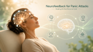
What Actually Helps: qEEG-Guided Neurofeedback for Post-Concussion Recovery
The Research Evidence
A 2014 PubMed study (NeuroRehabilitation) subjected two TBI patients to 20 sessions of EEG neurofeedback therapy. Cognitive scores and post-concussion symptoms improved significantly (p < 0.05), and — crucially — MRI showed significant increases in cortical grey matter volume and white matter fractional anisotropy following treatment. Neurofeedback was not just changing symptom scores; it was producing measurable neuroplastic changes in brain structure.
A 2025 VA Research RCT — the first to compare individualized infra-low frequency neurofeedback against a control group in Veterans with TBI — found that 20 sessions of personalized neurofeedback over 8–10 weeks significantly reduced chronic headaches, sleep problems, and attention disorders versus controls. The researchers concluded that individualised neurofeedback ‘holds promise to be a safe and effective intervention for those who suffer with post-concussive symptoms.’
A retrospective study of 40 PCS cases using qEEG-guided neurofeedback showed a significant reduction in post-concussion symptom severity, reduced slow and fast wave amplitude ratios, and normalisation of qEEG profiles. A separate case study found that 22 sessions of qEEG-guided neurofeedback produced complete cessation of headaches and full qEEG normalisation. A review across all TBI neurofeedback studies found that all 22 studies demonstrated positive findings across a wide range of neuropsychiatric symptoms.
How the Protocol Works at Bhakti
At Bhakti Brain Health Clinic, every post-concussion patient begins with a full qEEG brain mapping assessment that maps their specific dysregulation pattern — where excess delta and theta are concentrated, which regions show coherence disruption, and whether the Traumatic Brain Index shows a concussion-consistent profile. The neurofeedback protocol is then designed to directly target those findings: down-training excess slow-wave activity, restoring alpha rhythms, normalising high-beta hyperarousal, and retraining coherence between disconnected regions.
Progress is tracked with repeat qEEG assessments throughout the training course, providing objective data on which patterns are normalising. Where appropriate, HRV biofeedback is added to address the autonomic dysregulation — the chronic fight-or-flight activation — that frequently accompanies PCS, particularly in patients with comorbid PTSD or anxiety. Most patients engage in 20–40 sessions depending on injury severity and the complexity of their qEEG findings.
Frequently Asked Questions
Why do post-concussion symptoms last so long?
Post-concussion syndrome involves persistent brainwave dysregulation — particularly excess slow-wave delta and theta activity, suppressed alpha, and disrupted coherence between brain regions — that does not resolve through rest and time alone. The brain’s neurovascular coupling (resource distribution to active regions) fails to normalise after the acute injury window, leaving functionally impaired circuits that produce measurable abnormal electrical patterns. Standard MRI and CT cannot detect this because they image structure, not function. qEEG makes the underlying dysregulation visible and targetable.
Can qEEG detect post-concussion syndrome?
Yes. qEEG consistently identifies the brainwave abnormalities associated with post-concussion syndrome — excess waking delta, elevated theta, suppressed alpha, high-beta hyperarousal, theta/beta ratio elevation, and reduced coherence. Many qEEG databases also include Traumatic Brain Index (TBI Index) discriminant analysis, which compares a patient’s brainwave profile against known concussion patterns. This provides an objective, validated measure of concussion-consistent dysregulation that structural imaging cannot detect.
How many neurofeedback sessions does post-concussion recovery take?
Research protocols typically use 20–40 sessions over 8–12 weeks. The 2025 VA RCT used 20 sessions over 8–10 weeks (three 30-minute sessions per week). The 40-case qEEG-guided retrospective study showed significant symptom reduction within this range. More complex presentations with multiple symptom domains or longer injury histories may require more sessions. At Bhakti, the qEEG brain map guides the number of sessions needed, and progress is confirmed with repeat qEEG assessments.
Is neurofeedback safe for TBI and post-concussion patients?
Yes. Neurofeedback is non-invasive and drug-free — sensors only read brainwave activity, nothing is transmitted into the brain. It is particularly appropriate for TBI patients, for whom medication side effects can be more pronounced and whose brains are already managing elevated neurological stress. The 2025 VA RCT and all published TBI neurofeedback studies confirm a strong safety profile. At Bhakti, qEEG assessment before training begins also screens for occult epilepsy — a relevant safety step for any TBI patient.
Post-Concussion Treatment at Bhakti Brain Health Clinic — Edina, MN
Bhakti Brain Health Clinic is a specialist neurotherapy clinic in Edina, Minnesota, serving patients across the greater Minneapolis–Saint Paul area. Our qEEG-guided neurofeedback programme for post-concussion syndrome and traumatic brain injury is built on the same core principle as all our treatment: objective brain data first, personalised protocol second, measurable progress throughout.
If you are still experiencing headaches, brain fog, memory difficulties, mood changes, or sleep disruption months or years after a concussion — and have been told your brain scans look normal — a qEEG brain map at Bhakti may provide the neurological explanation your symptoms deserve. Contact us for a free 45-minute initial consultation.
Persistent Post-Concussion Symptoms? Start with a qEEG Brain Map.At Bhakti Brain Health Clinic in Edina, MN, our qEEG-guided neurofeedback programme is specifically designed to address the brainwave dysregulation that underlies post-concussion syndrome — drug-free, non-invasive, and personalised to your brain’s exact injury pattern. → Schedule Your Free Initial Consultation ← bhaktibrainhealthclinic.com • 888-783-BBHC (2242) • 7300 Metro Blvd #340, Edina, MN 55439 |
Post-concussion syndrome is not a psychological problem or a failure of willpower. It is a neurological condition — a pattern of brainwave dysregulation that persists after the structural injury has resolved. That means it is a brain state that can, in principle, be changed. qEEG makes the dysregulation visible. Neurofeedback provides the mechanism for retraining it. The research is consistent, the safety profile is strong, and the outcomes for patients who access qEEG-guided treatment are meaningfully better than rest and time alone. At Bhakti Brain Health Clinic, that is the treatment standard every post-concussion patient deserves access to.